Below are the questions/answers that were asked during the webinar. We will update this list as we start to answer them.
Q: Is JPEG2000 a lossy compression type? Is TIFF an appropriate alternative?
A: The JPEG2000 format offers lossless compression, at around a 2:1 ratio. It does offer much higher compression ratios (up to 20:1) as well that are lossy. We prefer JPEG2000 lossless compression over tiff because it is a streaming format which means you can load into memory portions of the image file. With a TIFF image you need to load the entire image at full resolution into memory. The larger 3D scan images that are very common today make this a cumbersome limitation. You can easily have an image that is hundreds tens or even hundreds of gigabytes in size when not compressed. Even if you have this much RAM in your workstation it will be incredibly slow to load as it transfers this image data off your disk to your RAM. With lossless JPEG2000 files you can load into memory the portion of the image you are currently viewing at the desired resolution (up to the maximum acquired resolution) further increasing the speed with which it opens. If you have additional questions please let me know.
Q: How to export an image with sholl analysis overlayed?
A: You can export your tracing with the sholl spheres/shells by using the following steps:
-
Open the file you would like to export
-
Perform Sholl analysis with the desired settings
-
Optionally you can perform any desired rotation
-
Go to the Publish tab/ribbon
-
Click the Image button in the EXPORT TRACING group
a. The icon is a blue rectangle with .tiff on it
-
Set you options and click Save File
a. Clicking this will bring you to the save dialog so you can name your file
Q: Does 360 have the capability to perform unbiased counting in any way? Or must this be done in Stereo Investigator?
A: Neurolucida and Neurolucida 360 do not perform unbiased counting. They can reconstruct and map, but for unbiased quantification/counting utilizing stereology you need to use Stereo Investigator.
Q: The marker tab don’t show all the markers, how can we make the tab smaller to view all the markers?
A: The best way to display the full marker tool bar is to undock it by clicking and dragging on the small gray horizontal line above the first marker. This will allow you to undock the marker bar and either tock it horizontally or leave it floating. Alternatively you can also minimize your ribbons by double clicking on the title of a ribbon such as Trace or Move. This will minimize or roll up the buttons on the ribbon giving you more vertical space on the screen.
Q: Can I export my plot with the markers (for example to Coreldraw) and still edit the whole plot (the line and the markers)?
A: You can export from Neurolucida into a vector graphics format by using the steps below:
- Open your file in Neurolucida
- Select the Publish tab/ribbon
- Click the Vector button in the EXPORT group
a. The help link below explains the options in this dialog
- From here you can export an .svg file that you can then open in vector graphics editors. I believe Corel Draw supports this format, but I’m not 100% sure.
Here is a link to the help article on the vector graphics export in our help: https://bit.ly/2YxUmJG
Q: Does Neurolucida360 include the basic Neurolucida?
A: Neurolucida 360 includes all the functionality of basic Neurolucida with the exception of the ability to connect to any hardware.
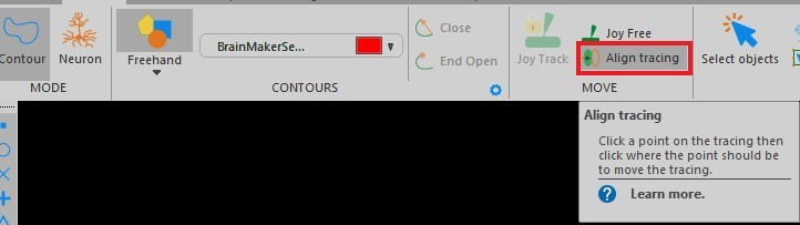
Q: If you need to pause during a job for some extended time, or if somehow the stage is bumped and the slide has been moved relative to objective, can you realign the sample and continue the tracing accurately?
A: If you have moved the slide or need to realign a tracing, you can go to the “align tracing” icon in the trace ribbon and select It. This will allow you to click on a point in the tracing, then the point where you want It to move to.
Q: Is it possible to outline cortical layers on the Neurolucida?
A: Yes, you can outline any regions utilizing contours, that you can distinguish in either your live camera image from your microscope or from images you have previously acquired on a different microscope.
You can also use open delineations to outline layers. Follow this link to our help article on how to create open delineations. You want the “Creating delineations: Procedure” section.
Q: I have also problems when I need to take a moisac of a tissue taking several photos of the tissue using a high objective.
A: Here are two help articles on slide scanning troubleshooting. The first is brightfield the second is fluorescent:
https://www.mbfbioscience.com/help/neurolucida/Default.htm#Imaging/SlideScan/SlideScan_TroubleshootBrightfield.htm?Highlight=Slide%20scanning:%20Troubleshooting%20
https://www.mbfbioscience.com/help/neurolucida/Default.htm#Imaging/SlideScan/SlideScan_TroubleshootFluo.htm
Q: Do image files need to be saved as a specific file type?
A: We support a wide variety of image formats. It depends on what you are doing. We typically recommend JPEG2000 (.jp2 or .jpx) because of its efficiency and speed. If you are going to another application with your images a .tif may be necessary as many other applications do not yet support JPEG2000. There are drawbacks with .tif images such as size restrictions and performance issues.
Q: Does spine analysis depend on species, rat/mouse/human ?
A: Spine analysis are not species specific. You can get spine data from any species or preparation as long as you can see the spines to then mark or trace them.