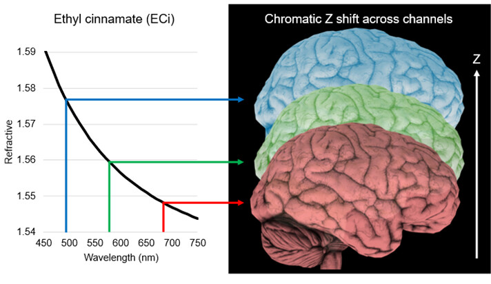
Chromatic Z-shift arises from the chromatic dispersion of the sample mounting medium.
Chromatic dispersion describes the phenomenon where a material’s refractive index varies with wavelength. When a mounting medium exhibits strong dispersion across the spectrum, fluorescence of different wavelengths propagates through the medium with slightly different optical paths. As a result, structures labeled with different fluorophores may be imaged at different depths even though they originate from the same physical location in the sample. This effect is noticeable when using hydrophobic mounting mediums like ethyl cinnamate (ECi). ECi is widely used because its refractive index closely matches that of cleared tissue, providing excellent transparency and deep tissue imaging capability. However, ECi exhibits strong chromatic dispersion, which can introduce noticeable focal shifts between fluorescent channels across the visible spectrum.
In contrast, dibenzyl ether (DBE) is a hydrophobic mounting medium that generally exhibits lower chromatic dispersion. Although detailed dispersion measurements for DBE are limited in the literature, practical imaging experience indicates that DBE-based mounting medium typically introduces only small chromatic Z shifts. In many cases, this minor shift can be compensated using the channel-specific refractive index compensation maps configured during the SLICE Tissue Scan Workflow to allow accurate colocalization analysis.
In summary, it is critical to consider not only the refractive index matching of a mounting medium but also its optical dispersion properties when planning multi-channel imaging experiments for light sheet microscopy. If colocalization analysis between fluorescent channels is part of the experimental design, clearing methods with lower chromatic dispersion, such as DBE-based solvent clearing or certain aqueous clearing protocols, are recommended.